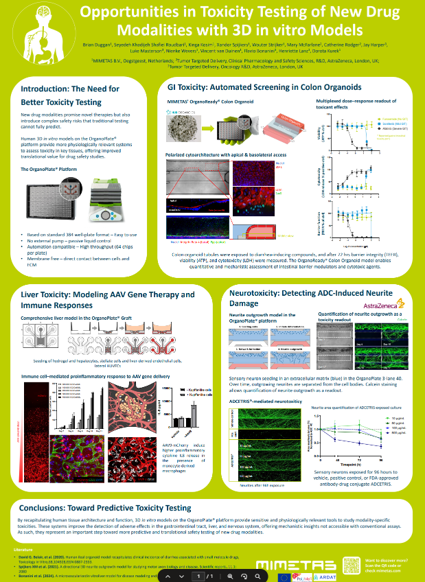
Assessing Vascular Inflammation and Safety with the Perfusable 3D OrganoReady® Blood Vessel HUVEC
.avif)

Why This Is Important

Challenges

Need
MIMETAS’ Answer
.svg)
Organ Model
A ready-to-use human 3D vascular model featuring 64 perfusable tubular structures per plate, enabling physiologically relevant inflammation, toxicity, and monocyte adhesion assays.
Features
• Perfusable lumen with controlled apical & basal access
• Stable, functional and leak tight endothelial barrier
• Reproducible inflammatory activation• Robust monocyte adhesion assays
• Reliable toxicity read-outs for screening
Offering
OrganoReady Blood Vessel HUVEC
Custom CRO Services
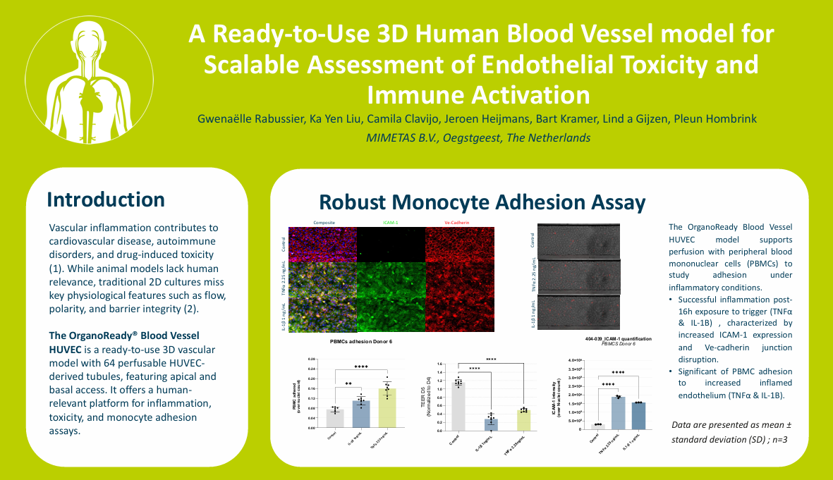
Studying Vascular Inflammation
Vascular inflammation and monocyte adhesion are key drivers in many diseases. Physiologically-relevant endothelial models areessential to evaluate inflammatory pathways, drug responses, and vasculartoxicity.
Characterization of the Endothelial Vessel
High-resolution imaging of the OrganoReady Blood Vessel HUVEC reveals a well-organized endothelial structure, marked by continuous VE-cadherin junctions and aligned actin fibers along the tubular wall. This architecture reflects a stable, cohesive barrier in the 3D perfusable vessel (figure 1).

Figure 1. A 3D fluorescent image of the OrganoReady Blood Vessel HUVEC, showing nuclei (blue), VE-cadherin (red) and Actin (yellow).
Assessing Inflammation and Toxicity
The model delivers a strong dose-dependent toxicity profile after doxorubicin treatment, captured through real-time transepithelial/transendothelial electrical resistance (TEER) measurement (Figure 2A). Cytokine treatment also triggers a significant, dose-dependent upregulation of ICAM-1, confirming robust and physiologically relevant endothelial activation (Figure 2B).

Figure 2. Doxorubicin induced a concentration-dependent decrease in barrier integrity, with higher doses causing a faster and more pronounced TEER decline, while control conditions remained stable.

Figure 3. ICAM-1 expression was quantified 48 h after exposure to LPS, TNF-α, TNF-α + IFN-γ, or doxorubicin at the indicated concentrations. ICAM-1 intensity was normalized to nuclei count. Inflammatory stimuli induced significant, dose-dependent ICAM-1 upregulation, with the strongest response observed for TNF-α + IFN-γ, while doxorubicin produced a more modest effect.
Robust Monocyte Adhesion Assay
The model supports perfusion with peripheral blood mononuclear cells (PBMCs) to study adhesion under inflammatory conditions (Figure 3). TNF-α stimulation produces a marked increase in monocyte attachment, demonstrating that OrganoReady HUVEC can effectively capture key inflammation-driven cellular interactions.

Figure 3. Significant increase of PBMC adhesion to inflamed endothelium (TNFα and IL-1B). Data are presented as mean ±standard deviation (SD) ; n=7-8
Summary
- Physiologically Relevant Vascular Inflammation: A perfusable 3D human blood vessel model with a stable, leak-tight endothelial barrier enables robust modeling of inflammatory activation, barrier disruption, and monocyte adhesion under flow.
- Predictive Safety and Toxicity Assessment: Real-time TEER measurements and dose-dependent endothelial responses provide sensitive, human-relevant readouts for vascular toxicity and drug-induced inflammation.
- Translational Immune–Endothelium Interactions: Controlled cytokine stimulation and PBMC perfusion support reproducible assessment of ICAM-1 upregulation and immune cell adhesion, bridging mechanistic insight with screening-ready throughput.
References
- Henein, M. Y., Vancheri, S.,Longo, G., & Vancheri, F. (2022). The role of inflammation incardiovascular disease. International journal of molecular sciences, 23(21),12906.
- Nam, U., Lee, S., Ahmad, A., Yi, H. G., &Jeon, J. S. (2024). Microphysiological systems as organ-specific in vitrovascular models for disease modeling. BioChip Journal, 18(3), 345-356.

Selected Resources
.avif)

