Metabolic Disease Drug Discovery
MIMETAS delivers advanced 3D tissue models that help researchers explore disease mechanisms and therapeutic strategies with confidence.
Our assays address key biological processes such as tissue damage, inflammation, fibrosis, and vascular dysfunction – all designed to deliver actionable insights across a spectrum of metabolic disorders.
.avif)
.avif)
The Most Relevant
Human Liver Model
Currently Available
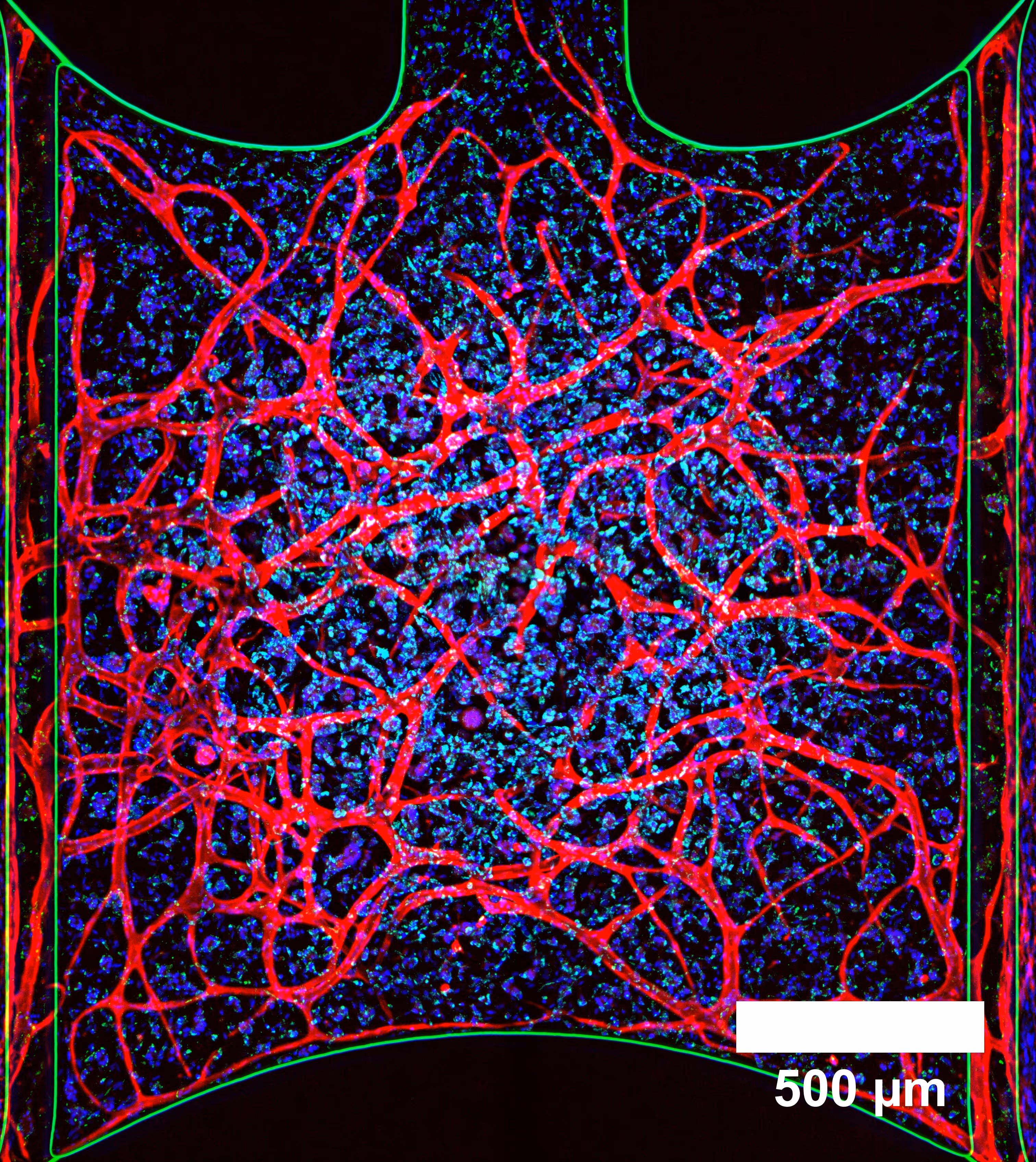
At the center of our metabolic disease portfolio is a vascularized, immune-competent liver model – developed to capture the liver microenvironment with an in vitro architecture that brings together all key cell types. This advanced system supports in-depth studies of conditions such as MAFLD and MASH within a controlled, physiologically relevant environment.
- Human hepatocytes (primary, iPSC-derived, or ASC-organoid-derived)
- Perfused LDEC vasculature
- Hepatic stellate cells
- Kupffer cells
- Circulating, perfused immune cells
With strong metabolic performance (e.g., albumin production, enzyme activity) and relevant pathological responses (e.g., IL-6, CRP, fibrosis), it serves as a powerful tool for exploring disease mechanisms and therapeutic strategies.
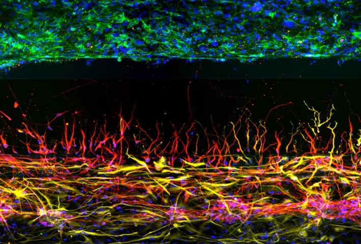
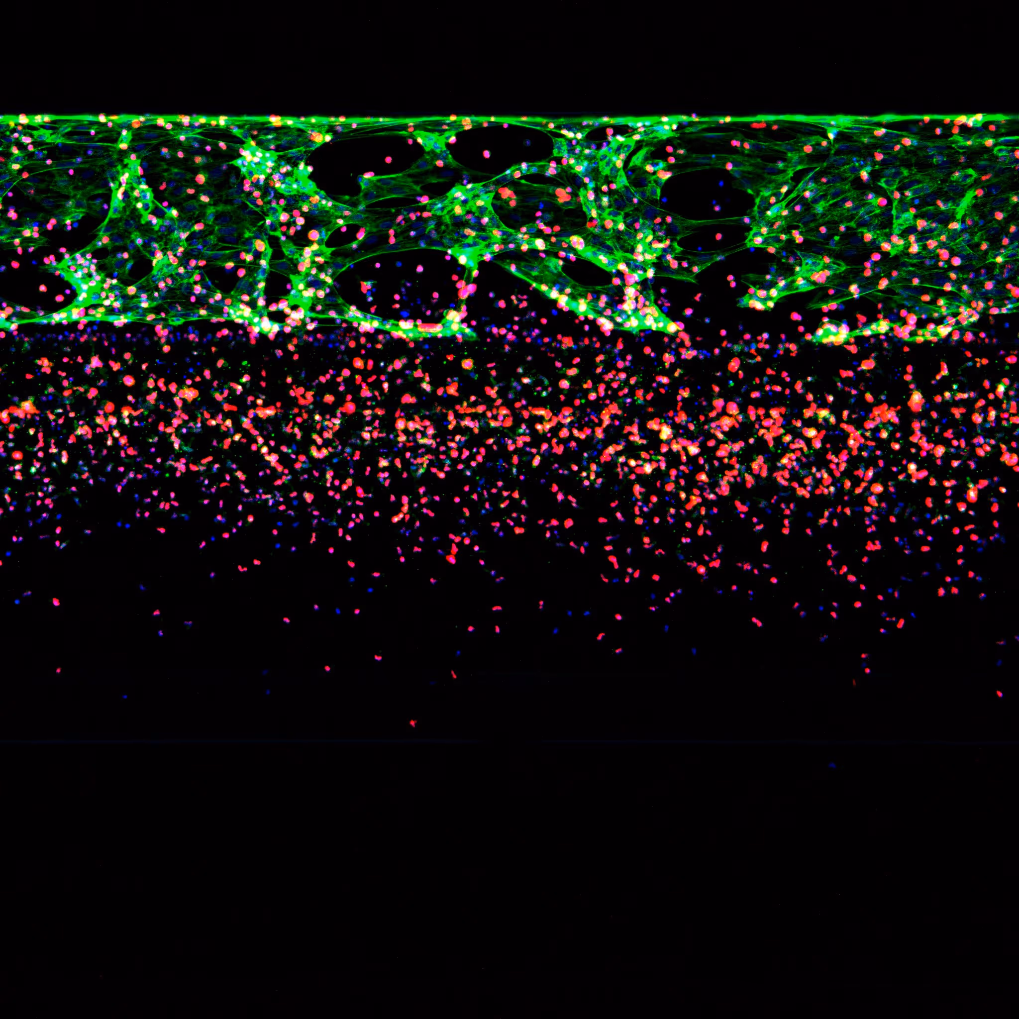
Assays Designed to Reflect Critical Processes in Metabolic Disease
MIMETAS researches metabolic diseases across organs of the liver, kidney, brain and vasculature. Our models capture essential features of metabolic disorders –providing platforms for target validation, mechanistic studies, and therapeutic evaluation.
Explore select applications below.
MAFLD: Steatosis and Lipid Uptake
.jpg)
MASH and Fibrosis: Screening for Anti-Fibrotic Compounds
Endothelial Dysfunction in a Coronary Artery Model
Vascular Inflammation
%20--%20these%20two%20images%20should%20be%20side%20by%20side.avif)