Microfluidic Culture Plates for Perfused 3D Cell Culture
Tools for creating dynamic, human-relevant tissue models.
The OrganoPlate® provides a reliable, scalable foundation for modeling in vivo-like biology, directly at the bench and on your terms.
Non-absorbant materials |Proprietary manufacturing
MPS Industry standard since 2014
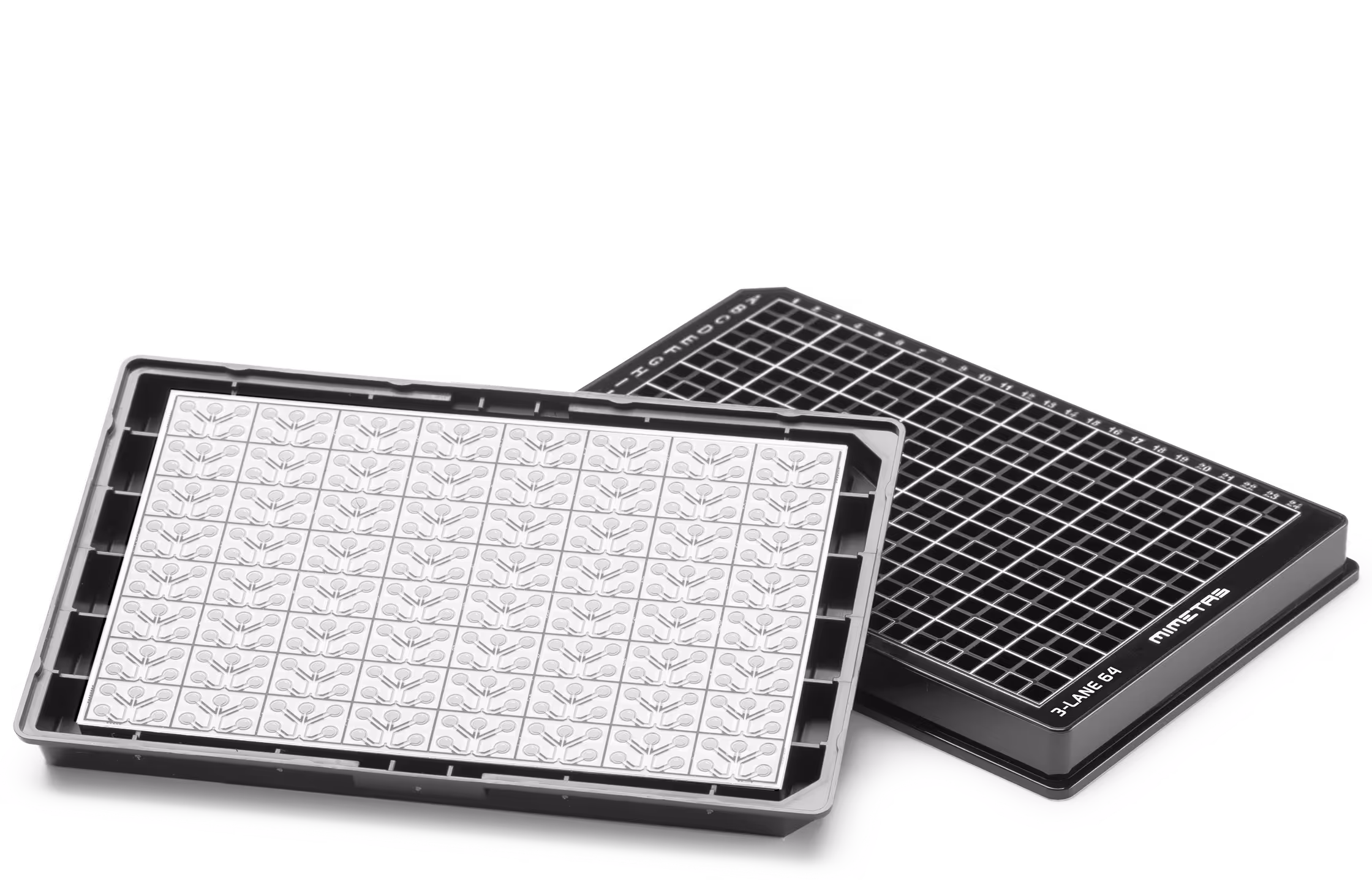
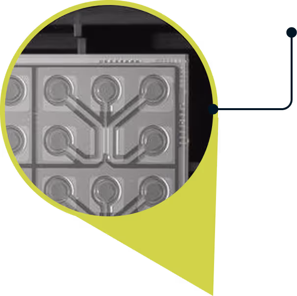
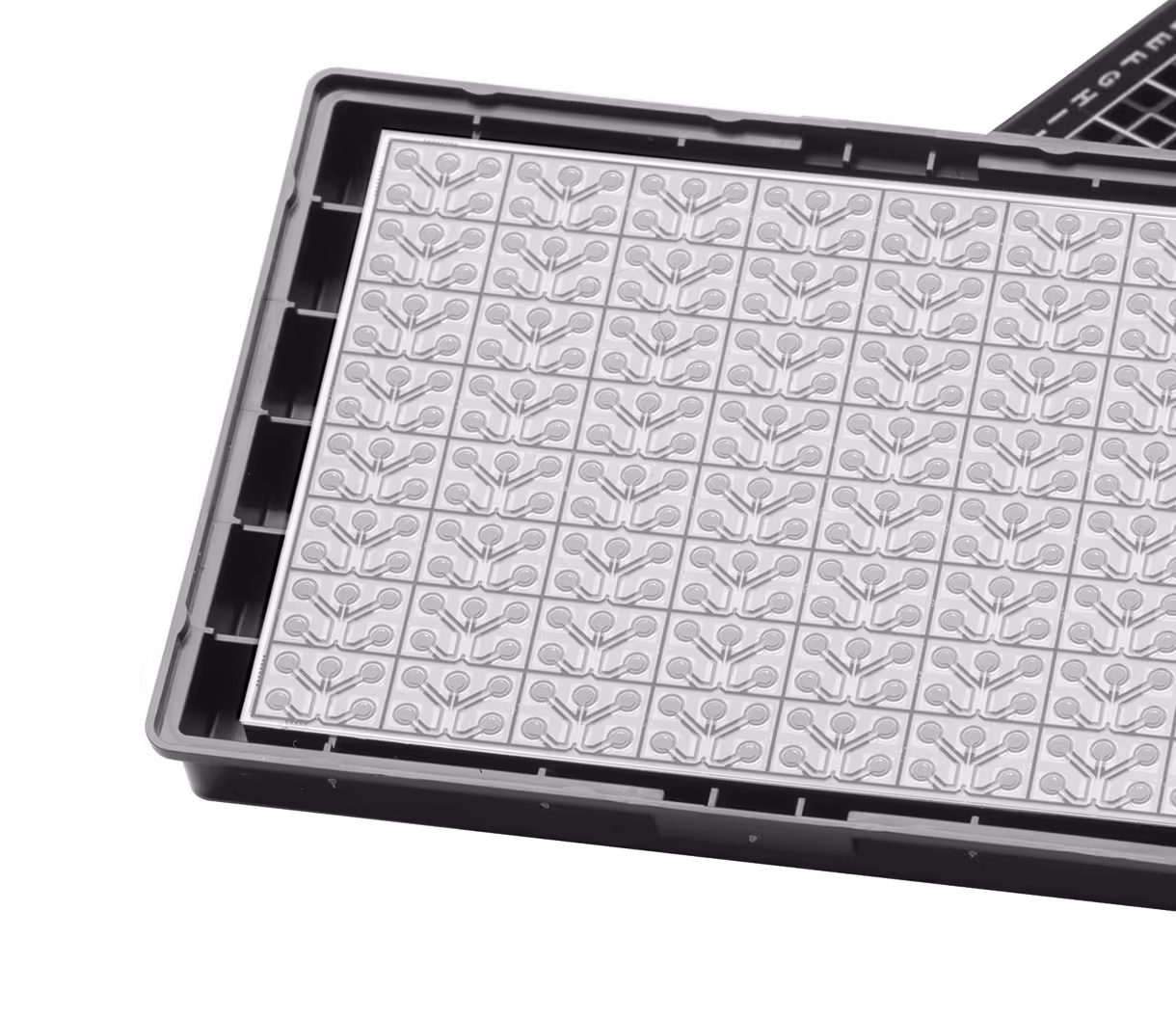
- 40 to 96 microfluidic chips
- 150μm thin microscope grade glass
- Chips with proprietory PhaseGuideTM technology
- Microtiter plate footprint
- Top print for user orientation
One Platform for All Tissue Cultures.
With the OrganoPlate® platform you can capture all cell culture modalities with one single platform.
Perfused barrier models
Angiogenesis and cellular outgrowth
Vascular network
Cell migration
Co-culture systems
Choose the Format That Fits Your Biology
| Format | Specification | Best for |
|---|---|---|
Format: OrganoPlate® 3-lane 64 | Specification: 64 tissue culture chips, 3 channels per chip, apical and basolateral access | Best for: Perfused epithelial tubules and blood vessels, co-culture, TEER and transport measurements, angiogenesis and cell migration Optimized for automation workflows and easy manual handling. |
Format: OrganoPlate® 3-lane 40 | Specification: 40 tissue culture chips, 3 channels per chip, apical and basolateral access | Best for: Perfused epithelial tubules and blood vessels, co-culture, TEER and transport measurements, angiogenesis and cell migration. |
Format: OrganoPlate® Graft | Specification: 64 tissue culture chips, 1 grafting chamber with 2 adjacent channels, apical and basolateral access | Best for: Tissue grafting (organoid, spheroid or tumor tissue) up to 1mm, perfused 3D culture, 3-layer co-culture, barrier integrity and transport, angiogenesis, gradient formation and cell migration. Optimized for automation workflows. |
Format: OrganoPlate® 2-lane 96 | Specification: 96 tissue culture chips, 2 channels per chip, apical access only | Best for: Perfused epithelial tubules and blood vessels, 2-layer co-culture. |
Getting Started With the OrganoPlate® Platform
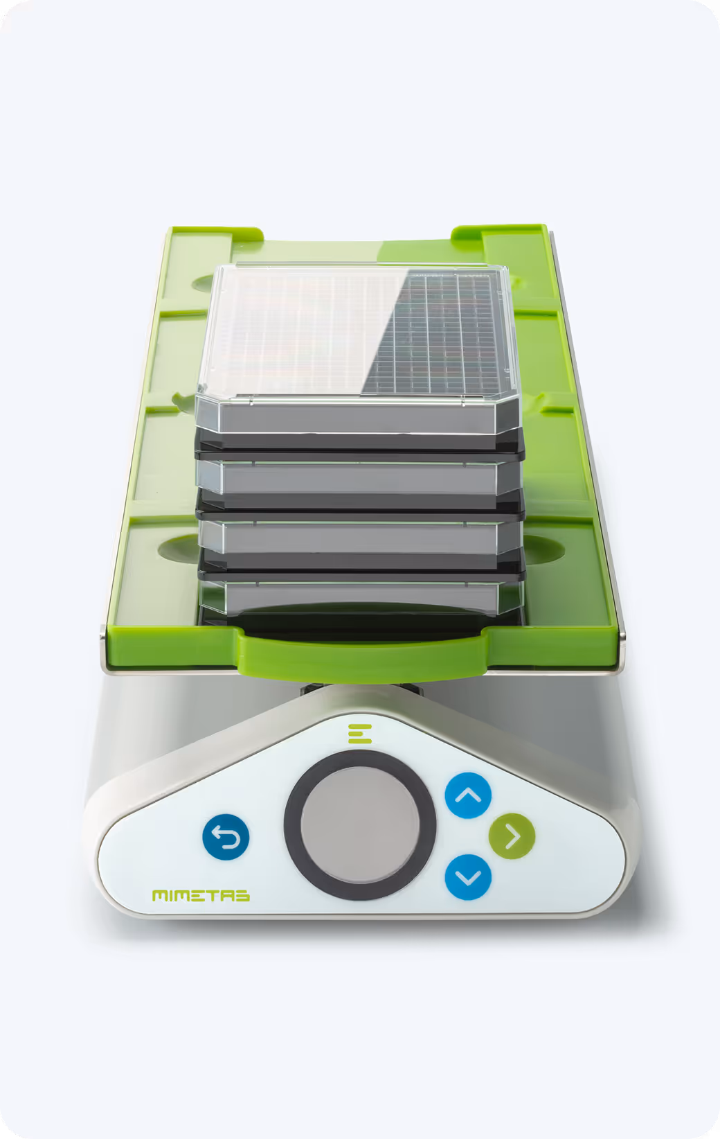
Our platform is designed with ease-of-use and flexibility in mind – all you need is an OrganoPlate®, a pipette and an OrganoFlow® device to get your 3D, perfused cultures started.
MIMETAS offers starter packages to help you quickly implement the OrganoPlate platform in your lab, including training on standard applications.
For more advanced users, we recommend the OrganoTEER®device, a reliable solution for real-time monitoring of epithelial barrier integrity, supporting both quality control and experimental readouts.
Looking to streamline your workflow even further? Our OrganoReady® models are pre-cultured and shipped assay-ready, allowing you to start compound testing immediately upon arrival.
Check out our offerings:
Learn about our custom CRO Services and see how our team can support your science in our labs.
Philip Hewitt
Early Investigative Toxicology,
Pre-clinical Safety, Merck

“The OrganoPlate® platform with the OrganoTEER® device will become a standardized assay in our early investigative toxicology drug screening workflow. With the OrganoPlate® platform, we see a reproducible early screen for intestinal toxicity, and the first example internally of an advanced cell model being implemented into routine testing.
To date, the OrganoReady® Caco-2 model is for us, at Merck, the most suitable platform offering at the same time scalability, speed, robustness, ease of handling, with low compound needs.”
Ready to Design Your Next Model?
The OrganoPlate® platform gives you the technical power to build advanced in vitro biology. Whether you’re designing the model yourself or looking to partner with MIMETAS to bring it to life faster, we’re here to help.