Predictive ADME & Toxicity Testing Services
MIMETAS delivers physiologically-relevant, 3D human tissue models at scale that provide reliable and early insights into compound safety and pharmacokinetics.
Human-Centric Models to Assess Drug Safety, Transport, and Tolerability
Our 3D tissue models provide human data to accelerate and de-risk programs for therapeutic modalities like small molecules, bi-specific antibodies, ADCs, and cell & gene therapy.
Explore some of our featured applications below.
Blood-Brain Barrier (BBB)
Built on primary brain microvascular endothelial cells, with optional co-culture of pericytes, astrocytes, and neurons, the BBB model supports studies on antibody transcytosis, small molecule transport, and barrier integrity.
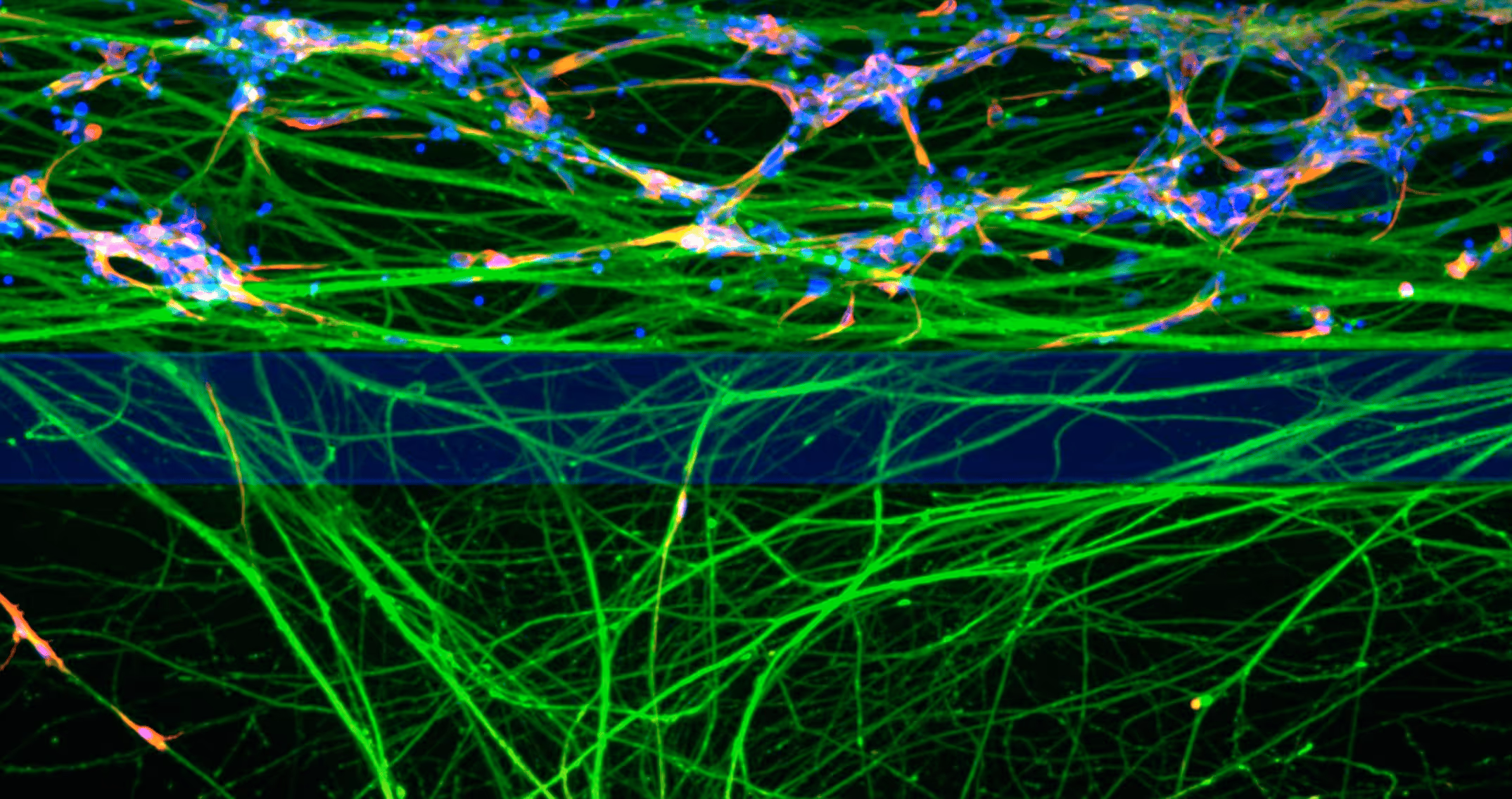
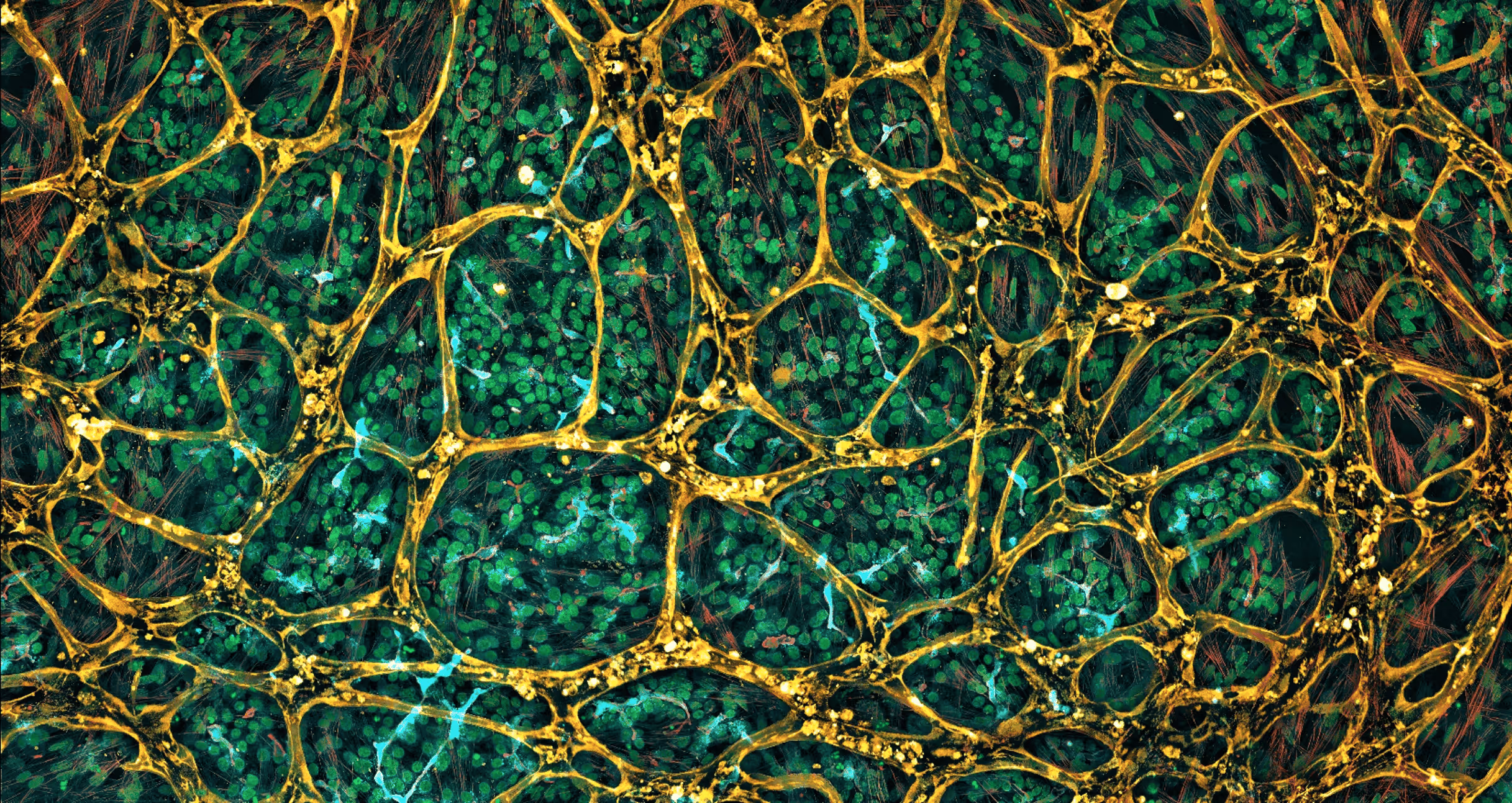
Chemotherapy-Induced Peripheral Neuropathy (CIPN)
Study neurotoxicity induced by chemotherapeutics and ADCs (Chemotherapy-Induced Peripheral Neuropathy - CIPN) and accurately identify safer alternatives by tracking axonal degeneration and cell health in human neurons.
.avif)
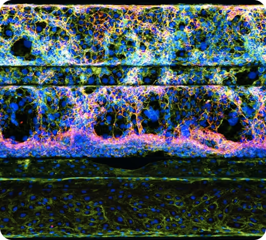
Gastrointestinal Toxicity
Our 3D human colon organoid model simulates dynamic epithelial and barrier integrity under flow, providing a reliable system for assessing GI toxicity in response to oral drugs and biologics.
Kidney
MIMETAS’ glomerular and proximal tubule, and ASC-derived organoids provide platforms for safety testing, mechanistic insights, and in-depth analysis of renal absorption, metabolism, and excretion.
Liver
MIMETAS’ liver model accurately replicates liver architecture, metabolism, and microenvironment, enabling studies on DILI/iDILI, gene therapy (LNP/AAV) delivery and safety, and liver metabolism.
.avif)
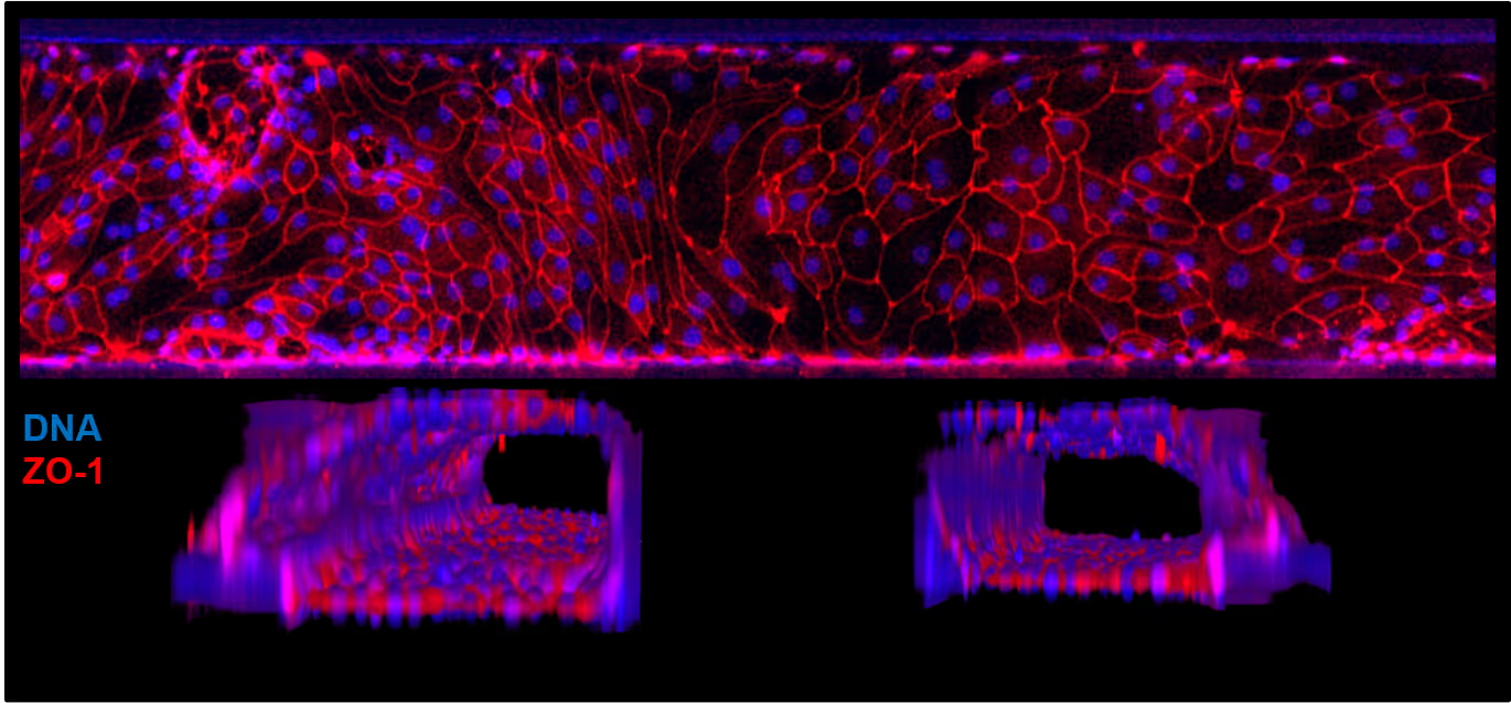
Vasculature
Perfusable endothelial tubules used for investigating vascular toxicity, assessing barrier function, marker expression and phenotypic analysis. Immune cells can be integrated to study vascular inflammation.
Built for High-Throughput, High-Impact Decision Making, At Scale
By recreating the complexity of human tissue environments, MIMETAS helps you evaluate drug safety and performance with confidence – before entering the clinic.
MIMETAS’ models enable human-relevant ADME/Tox studies by integrating:
Perfused endothelial and epithelial barriers
Organ-specific stromal and immune components
Functional outputs—cell viability, morphology, and permeability
Multiplex readouts for mechanistic insight