
MIMETAS and Wageningen University Researchers Demonstrate Real-Time Oxygen Consumption Monitoring in 3D Gut Models
Scientists at Wageningen University and MIMETAS have published a new study describing a method to monitor oxygen consumption in 3D gut-on-a-chip models. The approach enables researchers to assess mitochondrial respiration and follow compound-induced changes in cellular metabolism directly within the OrganoPlate® platform.
Published in Biotechnology Journal, the study “Liquid-Suspendable Metabolic Sensors Allow Real-Time Oxygen Consumption Monitoring in Caco-2 and Colon Organoid-Based Gut-on-a-Chip Models” demonstrates how oxygen-sensitive reagents can be used to generate functional metabolic readouts in both Caco-2 and adult stem cell-derived colon organoid tubules.
Studying Mitochondrial Function in 3D Tissue Models
Mitochondria play a central role in cellular energy production, and changes in their function are associated with intestinal disease and other chronic conditions. Measuring oxygen consumption provides insight into mitochondrial activity and how cells respond to experimental treatments.
However, obtaining these measurements in organ-on-a-chip systems can be challenging. Small fluid volumes and limited access to the tissue compartment complicate analysis, while existing methods often rely on embedded sensors or probes inserted into the device. The researchers therefore investigated whether a liquid-suspendable metabolic sensor could provide an oxygen consumption readout without modifying the chip.
An Oxygen Consumption Assay in the OrganoPlate
The workflow was established in the OrganoPlate 3-lane 40 using human intestinal epithelial Caco-2 tubules. An oxygen-sensitive reagent was added to the culture medium, and its signal was measured using a plate reader with time-resolved fluorescence detection.
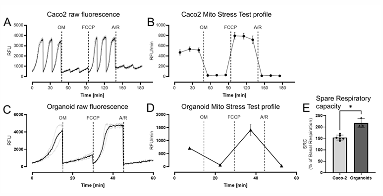
As cells consumed oxygen, the sensor signal increased, allowing changes in oxygen consumption to be followed over time. Measurements were performed during short periods without flow, separated by plate tilting to replenish oxygen in the microfluidic channels.
The researchers then sequentially added established mitochondrial respiration modulators to assess different components of cellular respiration.
Gut-on-a-Chip Models Show Measurable Respiratory Responses
Caco-2 tubules showed the expected responses to mitochondrial inhibition and uncoupling. Oligomycin reduced oxygen consumption, FCCP increased respiration following oligomycin treatment, and antimycin A combined with rotenone suppressed mitochondrial respiration.
These responses enabled the researchers to derive mitochondrial stress test profiles, providing information on basal, ATP-linked, and maximal respiration under the assay conditions, as well as proton leak and spare respiratory capacity.
The workflow was also applied to OrganoReady® Colon Caco-2 and OrganoReady® Colon Organoid models. Both models showed measurable changes in oxygen consumption following treatment. The colon organoid model exhibited a larger increase relative to its own baseline after FCCP exposure, although differences in cell composition and experimental workflows prevent a direct comparison of absolute respiratory capacity.
Expanding Functional Analysis in Gut-on-a-Chip Research
The study adds oxygen consumption monitoring to the functional readouts available for OrganoPlate-based gut models. By measuring cellular respiration directly within the microfluidic tissue environment, researchers can investigate metabolic responses alongside established approaches for assessing intestinal tissue function.
The method uses a sensor suspended in the culture medium, avoiding the need for embedded sensing components. Together, the findings establish an approach for mitochondrial stress testing in 3D intestinal models and provide a basis for further studies of intestinal metabolism and compound-induced metabolic changes.
This research was conducted within INIMINI-health, a public-private collaboration focused on developing intestine-on-a-chip models to study interactions between the intestinal epithelium, immune cells, and gut microbiota. The oxygen consumption assay adds a metabolic readout that supports this broader research effort.