

.svg)
Kidney NAMs: A scalable immunocompetent kidney-on-a-chip for function,inflammation, and drug response
Chronic kidney disease (CKD) and acute kidney injury (AKI) remain major global health burdens, yet translating preclinical kidney findings into the clinic is still notoriously difficult. Traditional models often struggle to capture human-relevant renal physiology, the dynamics of flow, and the immune-driven mechanisms that shape kidney injury and repair.
That's exactly where new approach methodologies (NAMs) come in. NAMs—human-relevant in vitro and in silico approaches—are designed to improve predictivity, reduce reliance on animal models, and enable mechanistic insight earlier in the drug development pipeline.
At MIMETAS, we've spent more than a decade building microfluidic kidney models on the OrganoPlate platform. Here's how our kidney-on-a-chip NAMs help researchers model renal function, disease biology, and drug response—at a scale that supports discovery.
Why kidney research needs NAMs that go beyond static culture
Kidneys are not "just" epithelial cells in a dish. Filtration, reabsorption, secretion, and inflammatory injury are shaped by:
- Polarized tubular architecture with distinct apical and basolateral compartments
- Barrier integrity (tight junctions) that governs selective permeability
- Transporter activity that drives drug handling and drug–drug interactions
- Vascular interfaces that influence oxygen/nutrient exchange and immune trafficking
- Immune cell infiltration and cytokine signaling that can accelerate damage and fibrosis
Static 2D models and many conventional systems miss one or more of these features—making it hard to study chronic processes, immune-mediated injury, or therapeutic interventions in a controlled, human-relevant way.
The MIMETAS kidney-on-a-chip approach (OrganoPlate®)
Our kidney models are built in the OrganoPlate microfluidic platform, based on a 384-well plate format that supports automation and high-throughput workflows. Within each chip, tissues are cultured against an extracellular matrix (ECM) gel under perfusion flow (generated by gravity-driven rocking), enabling more physiological cues than static culture.
Depending on the research question, the cellular complexity can be tuned—from epithelial-only tubules to co-cultures that include vasculature and immune components.
Key kidney models and what they enable
1) Proximal tubule-on-a-chip: function, transport, and nephrotoxicity
[NEDOSTAJE TEKST — screenshot je presečen ovde] … proximal tubule epithelial cells can form perfused tubular structures with measurable functional readouts.
What you can study:
- Barrier integrity and injury using fluorescent leakage assays, TEER measurements, and tight junction markers (e.g., ZO-1)
- Transporter-mediated uptake/efflux to assess renal clearance mechanisms and drug–drug interactions
- Drug-induced nephrotoxicity using multiparametric panels (cell damage, stress responses, functional readouts)
- Ischemia–reperfusion injury in co-culture formats, with protective-compound testing using apoptosis markers and TEER shifts
Together, these proximal tubule formats provide a strong foundation for functional and safety questions. When even greater physiological relevance is needed—especially to reflect donor variability—OrganoPlate kidney models can also be built using organoid-derived tissue sources.
2) Tubuloid-derived renal tubules: patient relevance with chip scalability
To increase physiological relevance, kidney tubule models can also be established from adult stem cell-derived kidney organoids ("tubuloids"). When dissociated and seeded into the OrganoPlate, tubuloids can form perfused, polarized tubules with access to both apical and basolateral compartments.
What you can study:
- Leak-tight barrier formation (fluorescent retention + TEER)
- Segment-relevant transport (e.g., functional transporter activity such as P-gp)
- Donor- and patient-derived variability as a foundation for stratification and personalized approaches
Because tubuloids can be derived from different donors, the model can capture donor- and patient-derived variability—creating a foundation for stratification approaches and, where relevant, more personalized study designs.
Spotlight: an immunocompetent kidney NAM for renal inflammation
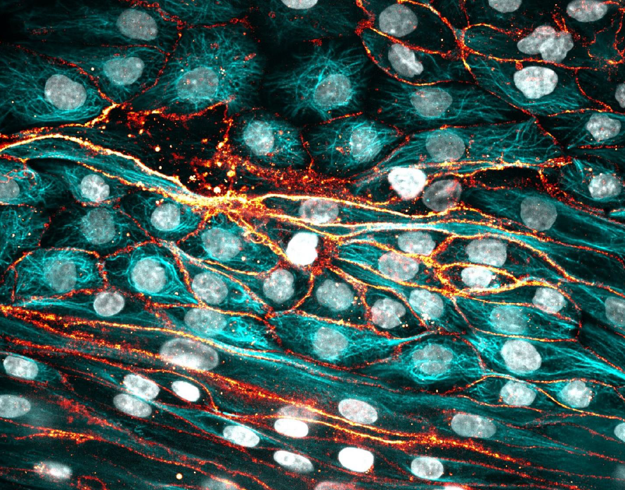
Many renal diseases are driven by immune dysregulation, but preclinical tools that capture immune–tissue crosstalk are limited. In a recent study, MIMETAS established a human immunocompetent proximal tubule-on-a-chip that recreates key aspects of the renal microenvironment: a perfused proximal tubule, a vascular interface, ECM, and primary immune cells.
While epithelial-only and tubuloid-derived tubules are powerful for function and transport questions, many kidney pathologies are driven—or amplified—by immune mechanisms. To address these questions in a controlled, human-relevant way, MIMETAS developed an immunocompetent kidney-on-a-chip format that incorporates vascular and immune components.
Below is what distinguishes this immunocompetent setup and what it enables researchers to measure.
What makes this model different
This model is differentiated by its ability to recreate immune–tissue crosstalk in a human, perfused kidney microenvironment while remaining scalable. It uses a membrane-free co-culture design in which renal epithelium and endothelium are cultured in direct contact with a collagen-I ECM, enabling physiologically relevant cell–matrix interactions. Primary human monocytes can be introduced under perfusion into the endothelial lumen, and inflammation can be triggered in a controlled way using complement activated serum (CAS). Because immune cell behavior can be monitored in real time, the system enables quantitative tracking of monocyte adhesion, extravasation, and migration over time, all in a format compatible with automation and quantitative assays.
With these components in place, the model captures both inflammatory signaling and functional immune cell behavior in the same assay window.
What the model shows (and why it matters)
Under CAS-triggered conditions, the system captures multiple hallmarks of renal inflammation and immune-mediated injury, including epithelial morphological changes, upregulation of inflammatory and adhesion markers such as ICAM-1, and increased release of pro-inflammatory cytokines such as IL-6. It also enables quantification of immune cell migration into the ECM and renal compartments, including donor-to-donor differences that are often masked in simpler assays. Importantly, the model supports therapeutic evaluation: immune-modulating compounds that act on either the inflammatory trigger or monocyte activity can significantly reduce monocyte migration into the renal compartment, demonstrating clear applicability for compound ranking and mechanism-informed screening.
These capabilities make the platform useful not only for mechanistic insight, but also for practical decision-making in drug discovery and development.
Where kidney-on-a-chip NAMs fit in the drug development workflow
Because OrganoPlate kidney models combine human relevance with scalable, quantitative readouts, they can support multiple stages of R&D. They are well suited for deciphering disease biology—especially mechanisms involving immune–tissue interactions—while also providing a human-relevant microenvironment for target validation. In discovery settings, they can be used for compound screening and ranking using multiparametric endpoints, and they also fit naturally into safety and ADME/tox workflows, including assessment of transporter-mediated effects that influence renal handling and toxicity risk.
How to leverage MIMETAS kidney capabilities
MIMETAS offers kidney NAMs in formats that match different needs:
- CRO Services for custom model and assay development, disease modeling, and compound testing
- Products for OrganoPlate-based solutions and tools to run models in-house (including ready-to-use options depending on application).
Takeaway: kidney NAMs that connect mechanism, predictivity, and scale
Kidney disease biology is complex—and often immune-driven. With OrganoPlate kidney-on-a-chip NAMs, researchers can move beyond static culture toward perfused, polarized, quantitative, and scalable kidney models that support both fundamental research and drug discovery.
If you're exploring kidney function, inflammation, or drug response—and want a NAM that can translate—MIMETAS' kidney-on-a-chip models offer a powerful next step.
Interested in the immunocompetent kidney model? Explore the case study and publication, or talk to a MIMETAS expert to discuss your research question and the best-fit model configuration.