Evaluate the effect of chemotactic triggers or cells on the migration of cells
Why 3D cell migration?
Cell migration is critical in establishing and maintaining human multicellular tissues. For example, wound healing and an immune response require proper movements of cells to specific locations, while aberrant cell migration can lead to various pathologies such as metastasis and vascular diseases. Cell migration is a multi-scale and multi-disciplinary process that depends on cell signaling, chemotaxis, and cell-matrix interactions. A quantitative 3D migration model in absence of artificial membranes with a stable gradient over time is essential to understand migration behavior in human tissues better.
We have optimized the OrganoPlate® platform to perform and study 3D cell migration. The excellent imaging quality allows you to see what happens in your tissue microenvironment, provides more read-out capabilities, and improves research quality. Evaluate 3D cell migration through an extracellular matrix (ECM) and maintain a stable gradient over time. Or study the effect of compounds or triggers on the mobilization and migration of cells. It's up to you.
Features
- Study immune, tumor, fibroblast and mesenchymal stem cell migration through an extracellular matrix
- No obstruction by artificial membranes
- Allows excellent imaging, cell tracing & quantification
- Stable chemotactic gradients over time
Assays
- Fluorescent barrier integrity assay
- Monocyte adhesion assay
- T cell migration assay
- Immunostaining for adhesion molecules
T cell extravasation from an endothelial vessel
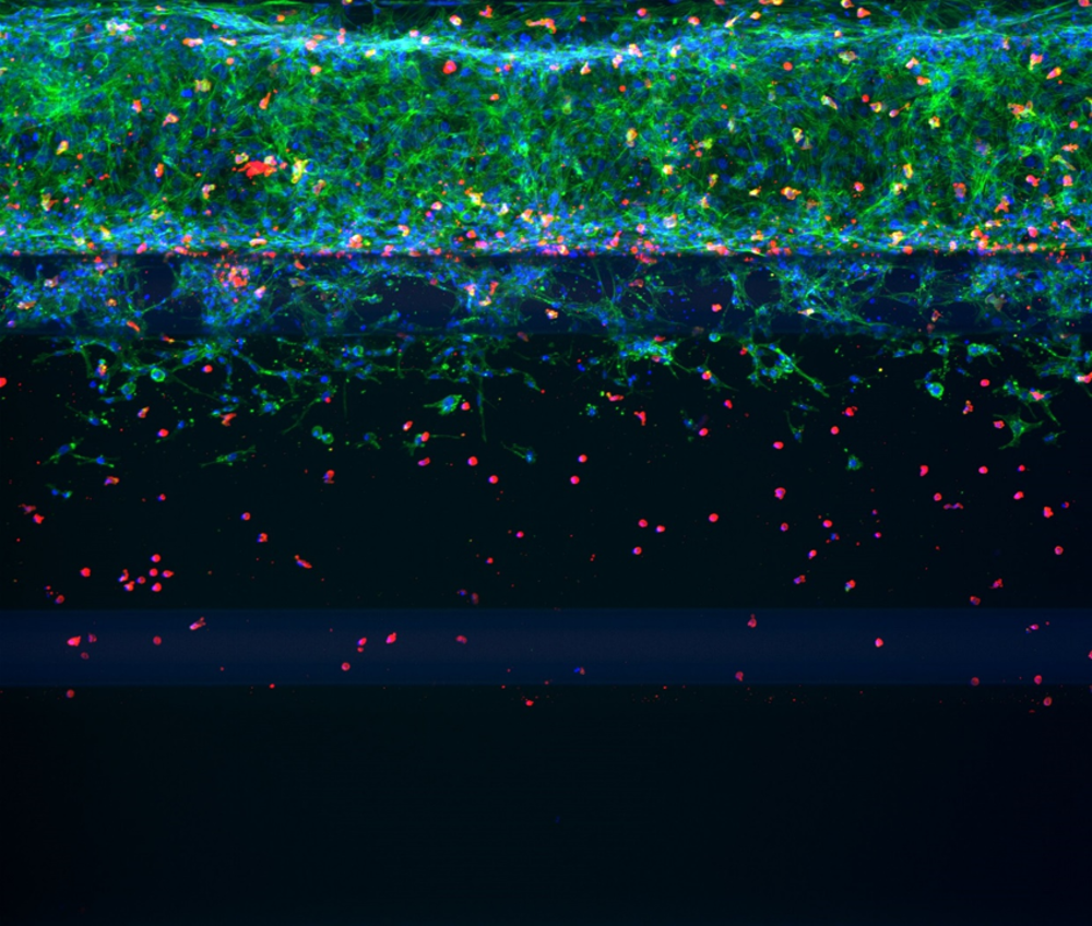
OrganoPlate® allows you to perform migration studies without the obstruction of artificial membranes. Study T cell migration by growing a perfused blood vessel, adding T cells, and the trigger of interest to eventually observe migration.
This example shows T cell extravasation from an endothelial vessel into collagen ECM in OrganoPlate® 3-lane 40.
T-cell marker CD45 (red) | Actin marker (green) | Nuclei marker DAPI (blue)

Want to know more?
Get up to speed with 3D tissue culture and learn how OrganoPlate® supports your research needs.

