 2D cell cultures have been used since the early 1900s. In recent years, 3D cell culture techniques have received much attention from scientists, as these might provide more accurate models of tissues.
Here, we will explore the differences between the two types of cell cultures and explain why 3D cell culture systems are becoming so popular.
A (Very) Brief History of 3D Cell Cultures
3D cell cultures have been around for longer than you might think. Ross Granville Harrison (1870–1959) adapted the hanging drop method from bacteriology to carry out the first tissue culture. This method allowed for further studies in embryology as well as experimental improvements in oncology, virology, genetics and a number of other fields.
The importance of the tissue microenvironment and 3D culturing techniques for cancer research was first proposed in the early 1980s by Mina Bissell, a lead researcher at Lawrence Berkeley National Laboratory. Since then, Mina has set up her own lab and continues to develop 3D techniques, especially in cancer discovery and treatment.
The 1990s were a quiet time for 3D cell cultures. This all started to change over the last five to 10 years. We’ve seen venture capitalists, angel investors and pharmaceuticals pouring lots of money into 3D cell culture technologies, research and production.
The reason? The enormous potential for 3D cell culture in drug development, which represents a big business opportunity. In our bodies, cells don’t grow in 2D, and it’s precisely the human body that we should model to develop better therapies against cancer and other diseases.
2D Cell Culture Systems
2D cell culture systems grow cells on flat dishes, typically made of plastic. The cells are put onto coated surfaces where they adhere and spread. Unfortunately, 2D cell cultures do have some inherent flaws:
- They aren’t representative of real cell environments — Growing on a flat surface isn’t a good way to understand how cells grow and function in a human body, where they surrounded by other cells in three dimensions.
- Lack of predictivity — 2D cell testing isn’t always predictive, which increases the cost and failure rate of new drug discovery and clinical trials. Pharmaceutical companies spend hundreds of millions on failed drug development every year.
- Issues caused by the growth media and expansion of cells — As cells grow in a standard 2D culture, they consume growth media and exude waste. This can result in toxic waste products, dead cells, nutrition depletion and damage of the environment the cells are in.
Why 2D Cultures Are Still Used in the Majority of Cell Research
Despite these disadvantages, 2D cell cultures are still used for the majority of cell cultures. Reasons include:
- It’s inexpensive — Economies of scale mean 2D cell culture systems and technologies are less expensive than some other systems.
- It’s well established — 2D cell cultures have been used since the early 1900s, gaining widespread acceptance in the 1940s and 1950s.
- There’s a lot of comparative literature — It’s easier to compare current results vs. previous results and studies.
- It’s what most researchers and scientists understand — Everyone involved with lab work has been taught 2D cell culture technology.
- Easier cell observation and measurement — 2D cell cultures are typically easier to analyze than some 3D cell culture systems.
3D Cell Culture Techniques
The reason for the growth in 3D cell culture is it’s simply a better way of representing human tissue outside the body. 2D cell cultures only exist in two dimensions. That’s not an accurate representation of how cells grow or how they are affected by disease and injury.
Although 2D cell cultures are still used for most research, the 3D cell culture industry is starting to catch up, especially in cancer treatment and stem cell research. Reasons for the increasing acceptance and use of 3D cell cultures include:
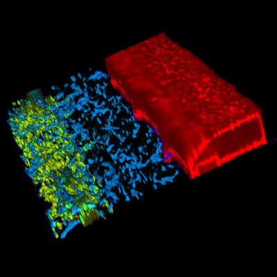
- More relevant cell models — Much better biomimetic tissue models make 3D cell cultures more physiologically relevant and predictive than 2D cultures. 3D plate cultures also show a higher degree of structural complexity and retain a “steady state” (homeostasis) for longer.
- Interaction between different types of cells — Creation of complex systems linked together by microfluidics means that 3D tissue systems can better model how different types of cells interact.
- Integration of flow — Fluid flow, for example blood flow, interstitial fluid flow and urine flow, is crucially important for the functioning of all tissues. Cells respond to flow through differentiation and metabolic adaptation
- Establishment of barrier tissues — Epithelia typically separate organ compartments, interact with the environment and protect the organism from the environment. Their proper functioning is crucial for survival and barrier malfunction plays a role in many diseases. Representation of barrier tissues is greatly enhanced in 3D culture.
- Better simulation of conditions in a living organism — Microfluidics continuously provide nutrients to where they’re needed, meaning cells and organs grow in a more realistic way.
- Reduces use of animal models — Animal models aren’t a reliable way to predict how drug treatments will affect humans. Screening drugs against human organs grown on chips is a much more reliable method.
- More realistic way to grow and treat tumor cells — 3D cell culture systems are good simulators of diseased tissue, including cancer tumors. They can exhibit similar growth and treatment patterns.
3D Cell Culture Techniques: Issues and Solutions
Some 3D cell culture systems do still have problems, but at Mimetas, we've come up with solutions.
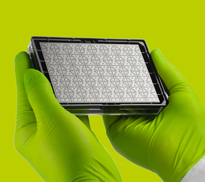
- Throughput — Many 3D culture techniques are cumbersome and time-consuming, rendering them unsuitable for drug development screening and research. The OrganoPlate® is based on a standard 384 well plate and provide the throughput needed for large-scale experimentation and screening.
- Challenges in microscopy and measurement — Due to the larger size of 3D cell cultures compared to 2D cell cultures, some types of measurement and microscopic analysis can be difficult. Mimetas has created technology specifically designed to allow for easy microscopy results, assays and readouts across all of its 3D plates.
- Getting oxygen and other essential nutrients to the right place — For larger cultures, it can be a challenge to distribute nutrients to where they need to be in the cell culture. Enhanced pump-free OrganoPlates®; vascularization in organ-on-a-chip devices, which Mimetas is developing; and architecture are helping to deal with this.
- Certain tissue models may contain undesirable elements — In rare cases, 3D cultures created from specific tissue types (e.g. basement membrane extracts) can contain unwanted components such as growth factors or viruses.
A Vital Part of 3D Cell Culture: The 3D Scaffold
There are two main types of 3D cell culture systems, a scaffold system, and a scaffold-free system. Scaffold-free systems typically use techniques such as “hanging drop templates,” “magnetic levitation,” and “magnetic 3D bioprinting.” Although there’s been some development in these fields, most of the focus is on regular 3D scaffold systems.
A 3D scaffold provides a way for cells to grow in three dimensions on a 3D plate. These are typically provided through something called hydrogel, an extracellular matrix (ECM) in which cells can survive, grow and proliferate. These ECMs normally have tiny pores that allow the passage of nutrients and gasses to give the cells the environment they need to thrive. Well-developed and selected ECMs also provide essential cues to cells, rendering them crucial for the establishment of physiologically relevant 3D tissue cultures.
This means 3D cell cultures with ECMs can be accurately grown and measured, providing an ideal environment for drug discovery and development and other types of research.
The Evolution of 3D Cell Culture Systems
3D cell cultures, systems, plates, and techniques have come a huge way over the last 10 years. Still, we’ve barely started. As physicians, scientists, and pharmaceutical companies learn the potential of 3D tissue culture, we’ll see more rapid innovation and wide-ranging applications.
At MIMETAS, we’re proud to be at the forefront, making personalized medicine and affordable drug screening a reality for everyone with OrganoPlates and OrganoPlate-based applications.